


The Pathologist
Monthly Roundup from The Pathologist // April 2024
Welcome back to your monthly roundup from The Pathologist, sharing the stories that you, our fantastic community, found most engaging in March!
This month, Eric Walk writes about The importance of AI fluency in the era of digital. Helen Bristow interviews a team of pathologists who have brought telepathology to low-resource settings. And we’re just getting started!
See you in May!
Download the April 2024 issue of The Pathologist - << CLICK HERE >>
See you next month!
The Power List 2024 nominations are officially open!
Nominate someone inspirational today!
It’s that time of year again when we ask you to take a moment in your day to put forth the name of the individual/s you know whose achievements and work in laboratory medicine has been breaking boundaries.
All you need to do is click the link below and fill in the short nomination form. Once the nomination window closes the entries will be scrutinised by our esteemed panel of judges who will carefully review each submission and select the most deserving candidates for inclusion in the Pathologist Power List 2024.
This year, we're excited to introduce five distinct categories, each highlighting different aspects of excellence in laboratory medicine:
- Destined for Excellence - Rising stars poised to make significant contributions.
- In the Wings - Non-pathologist medicine professionals going above and beyond.
- Heroes of Pathology - Problem-solvers aiding those in difficulty.
- Champions for Change - Drivers of environmental, social, and governance initiatives.
- Idols of Innovation - Researchers, inventors, and transformers pushing boundaries.
We aim to celebrate a diverse spectrum of professionals within our field, from early-career trailblazers to unsung heroes, disaster responders, environmental advocates, policy influencers, and beyond.
Join us in celebrating excellence and shaping the future of our profession! Nominate now!
Nominations close on 31st May 2024.
What Do You Want? – The Pathologist Reader Survey
As the year draws to a close we’re feeling introspective. And so, we’re keen to hear our valued readers’ (that’s you) thoughts on our 2023 coverage of everything pathology to help us shape our output for 2024.
The Pathologist exists to report on the research, innovations, personalities, and policies that shape pathology – but to do that we need your help. We’re here to spotlight the stories you care about, the stories that need to be told.
That means we need to hear your thoughts. We need all of your expert opinions on pathology and our place within it. Do you want to see more long reads? More interviews? What trends do you know are lurking in molecular pathology? What exciting developments are going on in the world of technology aren’t being discussed? What frustrates/inspires/excites/shocks you in this big, important field of ours?
Below you’ll find a questionnaire that gives you the chance to make your voice heard. Remember: the more insights you offer, the better we can serve the pathology community.
Here at The Pathologist, we are looking forward to an inspiring 2024 – filled with opportunities for personal and professional development.
The Pathologist team is constantly striving to enhance its offerings to better serve our community of pathologists and laboratory medicine professionals around the world. As we gear up to start 2024, we are embarking on a new offering that aims to enrich your professional journey: Email Courses. Email Courses are a series of educational modules (typically 6–8) delivered by email, free of charge, with a pre-determined frequency (for example, daily, biweekly, weekly).
We want to ensure that these courses align with your expectations and aspirations for professional development for the new year. Would you spare a few minutes of your time to share your thoughts through this brief survey?
Educational e-book: ‘Catch’ the latest in infectious disease!
A curated collection of the most important infectious disease stories
Interested in infection? Gripped by organisms? You’ll love our latest e-book.
In this interactive download, you’ll find coverage of the intense world of infection – everything from interviews with experts to ground-breaking research. Hear from researcher Craig MacLean on the role of agriculture, antibacterials, and resistances among host AMPs; an interview with Royce H. Johnson on the increasing prevalence of valley fever; a chat with author Jonathan Kennedy on his theory that germs have shaped history; and so much more, sponsored by LGC Clinical Diagnostics | SeraCare.
Celebrating the Powerful Profiles from ASCP, Revvity, and XiFin
Spotlighting iconic figures from the field: Industries are shaped by leaders – the movers, shakers, and positive disruptors. From all kinds of backgrounds, these people are fundamental to the technological and scientific breakthroughs that improve patient care. In “Powerful Profiles,” we’ll be spotlighting individuals from ASCP, Revvity, and XiFin who are crucial to the success of the industry’s leaders.
<< CLICK HERE FOR THE LATEST UPDATES >>
The Power List 2023: Storybook Edition
Welcome to The Pathologist Power List 2023, featuring the tales of 25 laboratory professionals – told in their own words
The 2023 iteration of our annual Power List is a veritable pathology anthology – a collection of topical, anatomical, and sometimes comical stories from the lab. After hundreds of submissions, our expert judging panel believed these final 25 tales to be symbolic of the amazing work that goes on day in, day out across the field – spanning multiple topics, subspecialties, and continents.
When brainstorming for a theme for this year’s Power List, we wanted to flip the idea on its head somewhat. We wanted to shake things up and get to the heart of the laboratory experience. And who would be better equipped for the task than the people who live and breathe pathology and laboratory medicine every day? And so, we asked for your tales from the lab – the highs, the lows, the unexpected twists. The final result signals that each and every person working in the lab has a unique story to tell.
Whether inspiring or interesting, heart-warming or funny (thank you, Mr Head-Holes), we believe that the Power List 2023: Storybook Edition has something for everyone – while shining a spotlight on the important work that you all do.
<< CLICK HERE FOR MORE INFORMATION >>
Interested in all things infectious disease? Then check out ID Transmission today!
ID Transmission brings you the latest research and innovations in the field of infectious diseases and tackles hard-hitting and thorny topics that many others shy away from. Their mission is to keep you informed, start conversations, and connect all disciplines and specialities working within this fast-paced area.
There are a number of ways to stay up to date with the latest from ID Transmission…

 The Foundation is very proud to announce the publication of a brand-new, absolutely FREE e-book explaining gross (macroscopic) description entitled "Opening Pandora's Box", by Drs. Paul Stromberg, Dr. Dan Rissi, Dr. Claudio Barros, and with minimal contribution (explained in the preface) by Bruce Williams.
 Human Diseases Models Morphological Phenotyping (MorphoPHEN) consists of an integrated and interdisciplinary Erasmus Mundus Joint Master. It is conceived to alleviate the real shortage of experts in morphological mouse phenotyping, which is needed to generate a positive impact of preclinical biomedicine and improve human translatability (from the bench to the bedside).
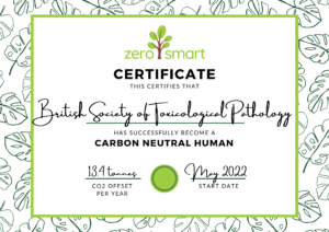 The BSTP is committed to sustainability in toxicological pathology and with the inclusion of the following paragraph in the constitution of the BSTP: